
| Researchers from the Laboratoire de Physique des Solides (Université Paris-Saclay/CNRS) and the Laboratoire Lumière, Matière et Interface (Université Paris-Saclay/ENS Paris-Saclay/CNRS/Centrale Supélec) have developed an experimental method to visualize in real time the growth of individual spherical viruses using total internal reflection fluorescence microscopy. |
All viruses on Earth exploit the cellular machinery of their host to replicate. One of the crucial steps in the life cycle of a virus involves a complex process of self-assembly, which occurs despite the macromolecular bulk and intense enzymatic activity prevailing in the cytoplasm. The simplest spherical viruses self-assemble by stochastic growth of their protein shell—called a capsid—around their genome consisting of one, or sometimes several, RNA chains.
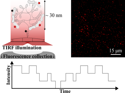
In this study, researchers from the Laboratoire de Physique des Solides and their collaborators implemented an experimental method to probe the growth dynamics of viruses at the level of individual proteins. The method is based on the use of total internal reflection fluorescence (TIRF) microscopy to follow the evolution of the light intensity of hundreds of assembly sites simultaneously as fluorescent proteins attach to or detach from them. Using a step detection algorithm combined with statistical analysis, the researchers were able to estimate microscopic quantities such as the attachment rate and the average residence time, which are inaccessible by traditional ensemble averaging techniques.
Single-molecule fluorescence imaging will prove crucial for elucidating virus self-assembly at the molecular level, particularly in crowded and active environments mimicking cytoplasmic conditions. These results are published in the scientific journal Nano Letters.
Contributors:
Équipe Structure et Dynamique d’Objets Biologiques Auto-Oganisés (SOBIO)
Laboratoire Lumière, Matière et Interface (LuMIn)
Centre Borelli
Funding:
LabEx PALM et NanoSaclay
References : T. Bugea, R. Suss, L. Gargowitsch, C. Truong, K. Perronet, G. Tresset, Nano Lett. 24 (2024) 14821-14828. https://doi.org/10.1021/acs.nanolett.4c04458
Contact :