Cryo electron microscopy for soft matter physics and interface physics-biology
Cryo electron microscopy (cryo-EM) let us visualize biological and soft matter objects at (sub) nanometer scale while preserving their hydrated state and native ionic environment, vitrified at low temperature. We access their conformations and interactions and are able to explore conformational changes, in the aim to relate these to their functional activities.
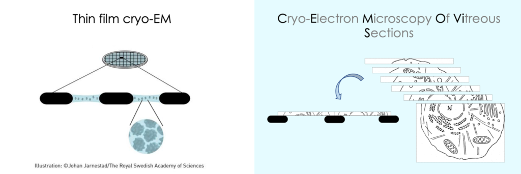
We use both thin film cryo-EM, to image macromolecular complexes and nano-objects (viruses, nanoparticles, liposomes, …) in solution, and CEMOVIS (Cryo-Electron Microscopy Of VItreous Sections) to image biological tissue and cells, or bulk liquid crystals and polymers.

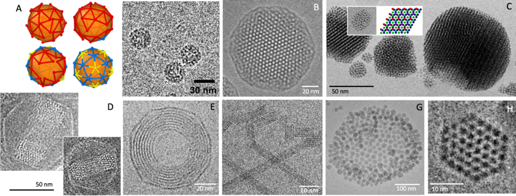
A few examples of thin film cryo-EM at LPS. (A) Viromimetic particles. (B) Cubosome (phytantriol/polymer/water). (C) Gold nanoparticle supra-crystals. (D, E) DNA toroids confined in bacteriophage (T5, Lambda) capsids. (F) Imogolite nanotubes. (G) Nano-emulsion stabilized by colloidal nanoparticles. (H) DNA fragments condensed by protamines.

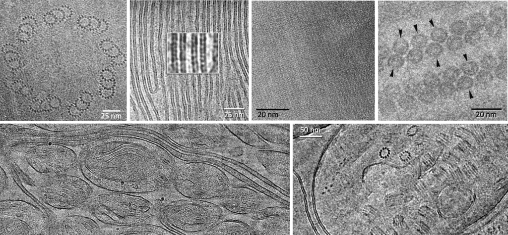
Cryo electron microscopy of vitreous sections. From left to right, top to bottom: basal body ofParamecium tetraurelia; thylakoid membranes of Euglena gracilis; intracellular crystalline inclusion; lamello-columnar phase of nucleosomes in solution; two views of Mushroom Bodies in Drosophila brain.

Equipments
- Cryo electron microscope JEOL 2010F: 200kV with field emission gun and CRYO pole piece, equipped with Hole Free Phase Plates and CCD camera (Ultrascan 4K, Gatan)
- Cryo-holders (Gatan 626) and cryo-transfer stations
- Thin film vitrification robots: Vitrobot Mark IV (Thermofisher) & homemade robot optimized for visquous specimens
- Liquid helium slam-freezing device for bulk specimen vitrification (Cryovacublock, Reichert)
- Cryo-ultramicrotome (Leica UC6/FC6) in controlled environment (T, RH), equipped with a micro-manipulator and fluorescence microscope
- Sputter/coater (Leica ACE)
- Plasma cleaner (Cressington)

The LPS cryo-EM platform is a plug-in-lab Paris-Saclay (CRYOTEM@LPS) and is part of the national facility METSA.
Contacts
Amélie Leforestier : amelie.leforestier@universite-paris-saclay.fr
Jéril Degrouard : jeril.degrouard@ universite-paris-saclay.fr
