
Epidemiological studies show a clear increase in renal lithiasis in most western countries, induced by changes in eating habits and lifestyle. Almost 20% of the population will be affected by this pathology during their lifetime. The majority of the kidney stones are of the idiopathic type and develop from pre-existing deposits of calcium phosphate — Randall’s plaques — whose formation mechanism is still poorly understood.
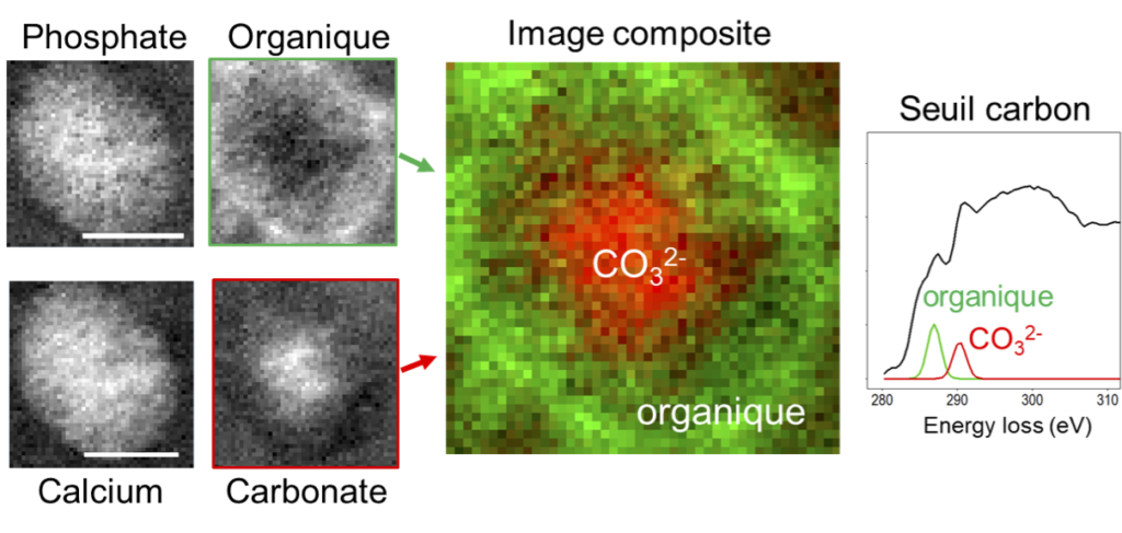
Researchers from the LPS and the LCPMC, in collaboration with medical doctors from Tenon Hospital, analysed samples of human kidneys to determine the location and composition of the nanometric-sized calcifications that correspond to the very early stages of Randall’s plaque formation. The characterisation of such objects within the renal tissue presents a daunting challenge due to their small size. The samples were analysed by electron energy-loss spectroscopy (EELS) in a STEM (scanning transmission electron microscope). The morphology and chemical composition were characterised at the nanometre scale for a large collection of objects, as was the interface between the organic and mineral phases. The results show that several types of nano-calcifications coexist. Despite certain specificities, the objects detected have striking similarities with those observed during the formation of physiological biominerals such as bone, and also with pathological calcifications associated with certain cardiovascular diseases. For instance, a significant fraction of the calcifications consists of calcium phosphate with a carbonate core surrounded by organic material (see figure). These similarities suggest common mechanisms between these different systems.
The strategy used in this work is of great interest, since it introduces a tool for the systematic exploration of pathological biominerals. Indeed, quantitative data are lacking for these systems and their complexity makes it difficult to propose models for their formation. Our study shows that STEM-EELS spectromicroscopy provides quantitative data allowing a reliable comparison with other more widely studied and better understood systems.
Reference
Nanoscale Analysis of Randall’s Plaques by Electron Energy Loss Spectromicroscopy: Insight in Early Biomineral Formation in Human Kidney
Clément Gay, Emmanuel Letavernier, Marie-Christine Verpont, Michael Walls, Dominique Bazin, Michel Daudon, Nadine Nassif, Odile Stephan, Marta de Frutos
ACS Nano 14, 2, 1823-36 (2020)
doi:10.1021/acsnano.9b07664